Written by The Mineralogical Research Group at the University of British Columbia
Special Thanks to Professor Lee A. Groat and Ph.D. student Catriona Breasley for this article
The Mineralogical Research Group at the University of British Columbia, under the leadership of Professor Lee A. Groat recently took delivery of a Leica DM750P microscope from Opti-Tech Scientific Inc. Professor Groat reports that the new instrument has transformed the work of the graduate students in the Group.
For example, Ph.D. student Lindsey Abdale studies climbing chalk with the new microscope. She is pleased to observe that the excellent resolution of the new instrument has made it much easier to find and identify minor impurities in commercially available climbing chalk.
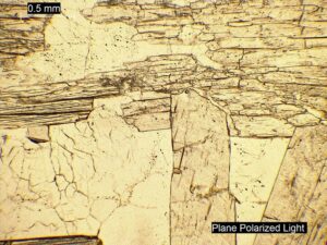
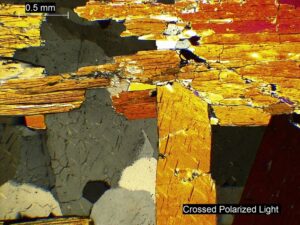
The research conducted by Ph.D. student Catriona Breasley begins with thin-section petrography; therefore, having access to a reliable microscope is invaluable. The high-quality camera attachment and colour resolution of the Leica DM750P allow for the capture of professional-quality images for use in academic publications, such as those shown below. Catriona would not hesitate to recommend a Leica microscope from Opti-Tech Scientific Inc. to any academic or industry professional looking for a top-of-the-range instrument.
Mary Macquistan’s M.Sc. thesis focusses on the characterization of a rare barium silicate mineral assemblage and relies upon accurate thin-section descriptions and high-resolution images of mineral relationships. The Leica DM750P outshines all microscopes she has previously worked with, and Mary highly recommends this instrument to any professional who does thin-section petrography. She has included a crossed-polar image of fresnoite from one of her thin sections to show the colour intensity and sharpness when looking through the eyepiece.
M.Sc. student Tiera Naber writes that thin section petrography is essential to her research on a rare earth element occurrence. The camera attachment on the Leica DM750P microscope enables her to capture high-resolution images of her rock samples to communicate her research to specialists and the general public accurately. Tiera notes that a quality picture truly is worth a thousand words.
The students, as is their supervisor, are very happy with the new microscope.

Catriona Breasley at the new microscope.


Catriona Breasley’s images of spodumene and quartz (plane-polarized above and cross-polarized below).


Plane-polarized (top) and cross-polarized (bottom; FOV 2 mm) images of fresnoite from Mary Macquistan.


Tiera Naber’s images of aegirine fenite (plane-polarized; FOV 3 mm) and amphibole biotite fenite (crossed polars; FOV 2 mm).
