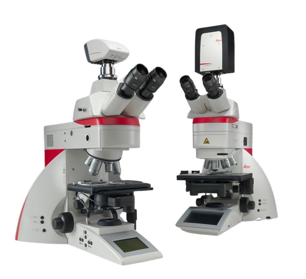
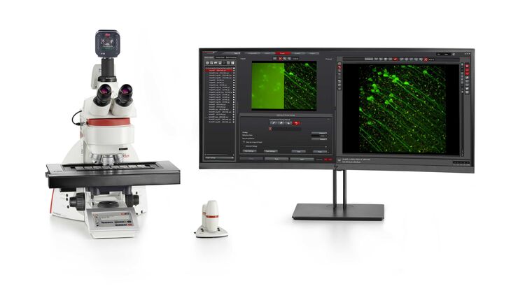
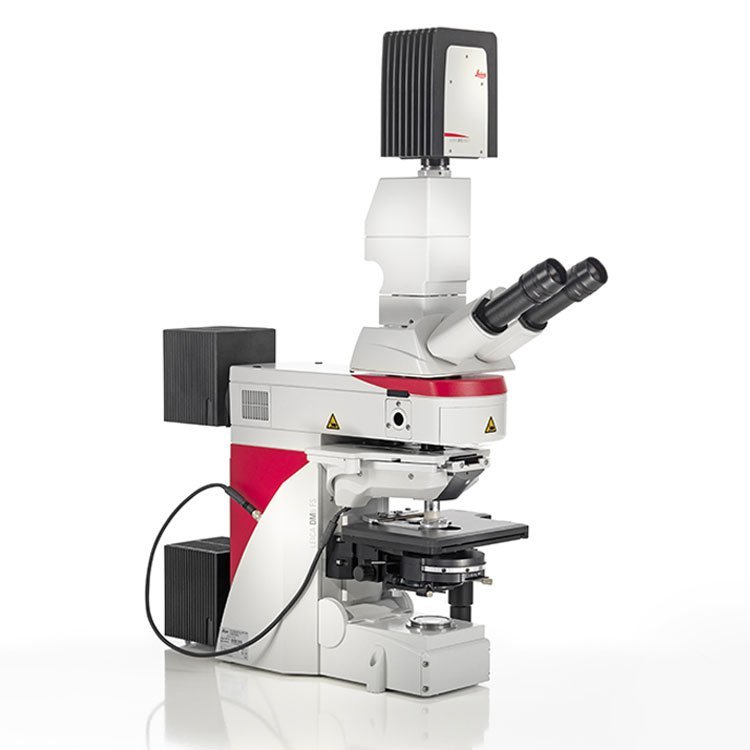
Looking to enhance your work efficiency? The Leica DM4 B and Leica DM6 B upright digital research microscopes are perfect for streamlining your work in biomedical research and clinical labs.
- Simplify your workflow with automated functions and easy-to-use software
- Easily capture publication-quality images by utilizing the 19-mm sCMOS camera imaging port
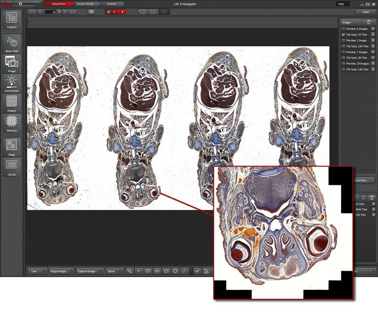
- Create fast overviews of your samples and identify the important details instantly with the LAS X Navigator Software
- Have great flexibility through the choice of accessories
Discover More in Less Time
By optimizing your image acquisition process, you can free up more time to focus on the core of your research. Here are some tips to help you streamline your workflow and increase efficiency.
- See up to 10,000 more of your sample with the LAS X Navigator. Set up high-resolution image acquisition for slides, dishes and multiwell plates
- Take full advantage of your sCMOS camera, for example, the Leica DFC9000! The 19-mm field of view camera port perfectly fits the dimensions of common sCMOS sensors.
- Make your slide examination faster at the highest resolution.
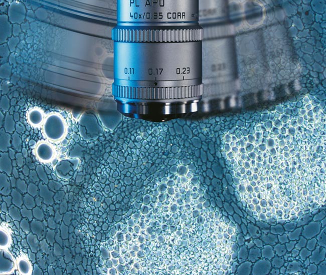
- Choose the objective that best fits your application – from more than 300 outstanding optics. One example is the unique 1.25x overview objective for a perfect wide view.
Free Your Mind!
Concentrate on the outcome you want to achieve instead of worrying about the steps to reach your goal. With Intelligent Automation, the hard work is done – multi-step routines are solved with a simple button press. This makes your work life easier, saves precious time, and frees you up to focus on your experiment rather than complex microscope settings.
- Change contrast with the push of a button – everything you need automatically slides into place.
- Acquire publication-ready images.
- Use Light and Contrast Management to simplify your work – for example, the Fluorescence Intensity Manager (FIM), fast Internal Filter wheel (IFW) and Excitation Manager (ExMan) for fluorescence images, or Koehler light management for perfect Koehler images.
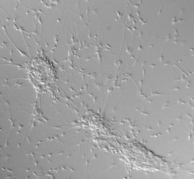
Contrast method DIC
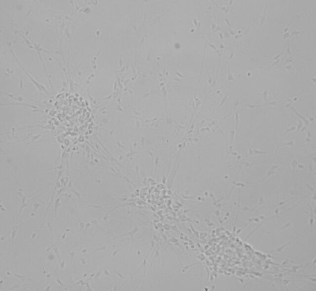
Unstained biological specimens often do not show a very high contrast.
Thick specimens, particularly brain slices, show up as nothing more than light grey structures instead of single cells.
Differential interference contrast
Differential interference contrast (DIC) is an ideal contrast method.
Exactly What You Need
With the Leica DM4 B and Leica DM6 B, you can customize your microscope to suit your
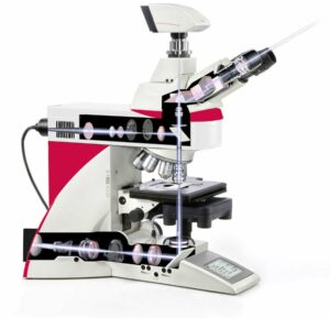
specific requirements and budget. Take advantage of the various contrast methods and automated functions to optimize your system for your unique application. The LED illumination guarantees uniform lighting for your sample with consistent colour temperature. Additionally, it is energy-efficient and eliminates the need for frequent bulb replacements, as it has a lifespan of up to 25,000 hours.
Make Your Microscope an Imaging System
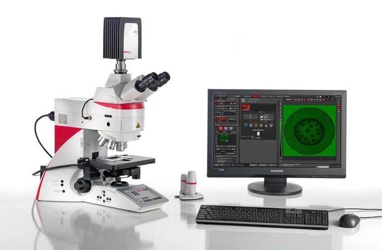
Leica Microsystems provides powerful cameras and workflow-based software to complete your integrated imaging system.
- Leica Application Suite X (LAS X) is our easy-to-use workflow-based software that guides you to acquire the images you need.
- Leica microscope cameras cover a broad range of applications from brightfield to fluorescence applications – or even one camera for multi-purposes such as our Leica DFC7000 T camera


