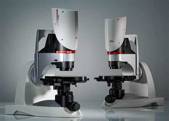
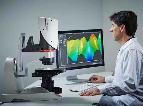
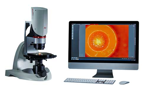
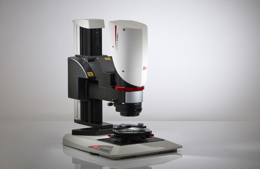
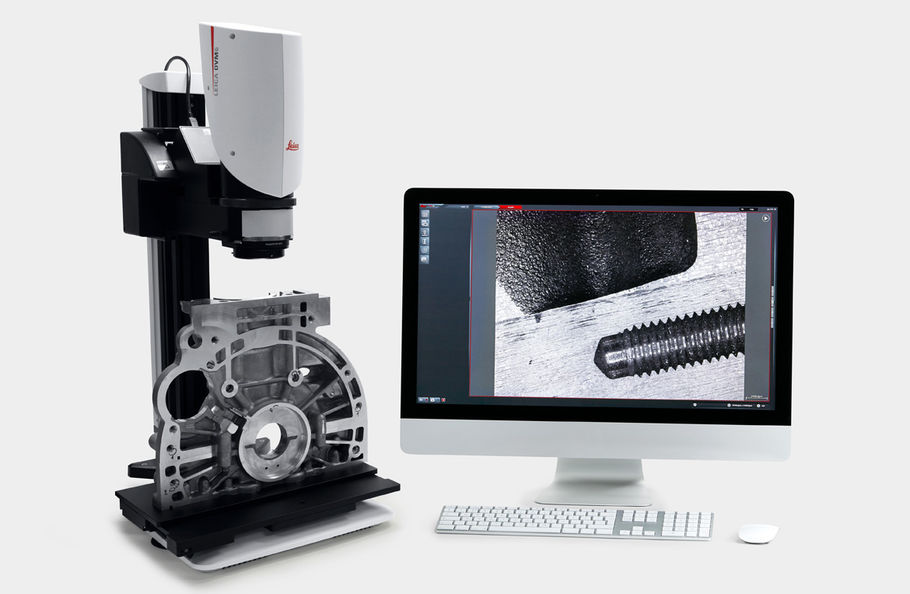
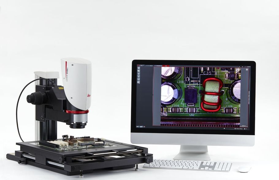
Digital Microscope Leica DVM6
Conducting precise failure analysis and research-and-development tasks can be challenging. However, the DVM6 digital microscope simplifies the process through its convenient operation and reliable results with reproducible imaging conditions, allowing you to stay focused on your work.
Go from macro to micro!
- 16:1 zoom range
- Three parfocal PlanApo objectives
- See details as small as 0.4 micrometres
- Change an objective with one hand
Low magnification objective: Ideally suited for large overviews which help you search and find efficiently regions of interest on your sample. It provides a max. field of view (FOV) of 43.75 mm, a long working distance up to 60 mm, and magnifications up to 190x.
Mid magnification objective: Gain more insights quickly and easily for your everyday work. It features a max. FOV of 12.55 mm, a comfortable working distance up to 33 mm, and a magnification range of 46x to 675x.
High magnification objective: See more fine details of your sample, down to 425 nm. It is capable of a max. FOV of 3.60 mm and a magnification up to 2,350x.
Large or bulky sample? Try the modular DVM6 M!
- Special interface adapter
- Zoom module can be used with Leica M series selected stands for:
- Large – max. stage travel range: 150 x 100 mm
- Tall – up to 340 mm
Try a different angle!
Observe your sample from -60° to +60° angle with the DVM6 tilting function. You can explore your sample from different perspectives by turning and shifting the stage to reach any point within a 70mm x 50mm travel range. The best part is that you can change this perspective with one hand, and the sample always stays focused since the tilting axis is aligned with the focus point.
Light and Contrast Combinations
- View samples with a textured surface using one or all four segments of the ring light illumination (RL)
- Visualise flat, reflective samples with coaxial illumination (CXI)
- Explore hard-to-image regions, e.g. holes and transparent materials, with the transmitted light illumination (BLI)
- Reduce glare on reflective samples, such as polished metal surfaces, with the diffusor adapter
- Identify contamination due to its different material properties with the polarizer adapter
Rely on your results!
With reproducible imaging conditions, you can reproduce an entire image, encode functions to ensure every user achieves comparable results, and sensor-detected and encoded objectives save you time and effort. All systems settings are saved with the image data and can be recalled anytime to compare your results.


