
The story of microscopy begins as a novelty item. The microscope, which was originally used to magnify small insects, later evolved into the first compound microscope in 1590, created by Dutch father and son spectacle-makers, Hans and Zacharias Janssen.

In the mid-seventeenth century, the role of the microscope grew from just making things appear bigger to actually being used for scientific discoveries, thanks to tradesman Anthony Van Leeuwenhoek. Van Leeuwenhoek crafted his own 300X magnification lenses, which helped him make a wide variety of discoveries such as bacteria, nematodes, and spermatozoa.
With the advancement of technology in the 18th century, the use of microscopes became more common among scientists, due to the discovery that the combination of two types of glass decreased the chromatic effect.
The middle of the 19th century produced more advanced microscopes, similar to the ones used today. During these years, scientists solved various obstacles with the use of microscopes.
The next big advancement didn’t happen until 1965 when the first scanning electron microscope revolutionized the science world.
The Latest Innovations in Microscope Technologies

Since first invented, the microscope has become a universal tool in biology labs (and in many other applications).
The automation today’s world demands makes an advanced microscope an instrument capable of accessing all measures of modern biology, from single images to time-lapse recordings of anything from individual particles to whole organisms.
Below are four of the latest advancements in microscopy:
- The Structure of Amyloid Proteins
An optical microscopy technique was introduced by academics at Washington University, unveiling nanoscale features of misfolded amyloid proteins in the brain, a groundbreaking discovery in the research of Alzheimer’s and Parkinson’s disease.
- Fibre Laser Microscopy
Both the University of Hong Kong and Bielefeld University in Germany have found a way of producing far less noise than usual when using a fibre laser by creating a firm, compact version of the fibre laser.
- Nanoscale Microscopy
Research from the University of Illinois, has produced a tool solving challenges across many domains, giving scientists a direct view of nanoscale structures without doing any image post-processing.
- Nanoscale Chemical Imaging
The Beckman Institute for Advanced Science and Technology at the University of Illinois has uncovered a new technique enhancing the observation of nanoscale chemical imaging by reducing the noise and increasing the scope and precision of specimens for them to be optimally studied.
The Future of Microscopy Products
The goal of microscopy product manufacturers is to develop solutions to extract as much data as possible from lab samples.
Confocal laser scanning microscopy, an optical imaging technique for increasing definition, is the current standard used to highlight the 3D features of a sample, together with the latest technologies, researchers are able to get as much information as possible from images.
The future of microscopy lies in the development of fully digitized systems facilitating users to confidently view samples in 3D, with an unmatched look throughout a biological sample, and in the process, yielding accurate and reliable results.
Why Choose Opti-Tech
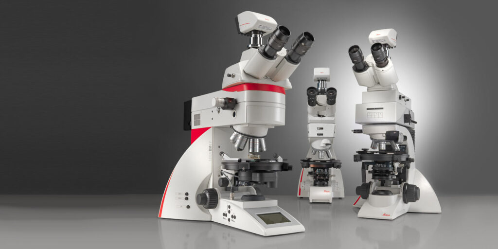
As a leading supplier of scientific equipment, specializing in optical & digital Microscopy, Opti-Tech provides uncompromising support from a sales team with unsurpassed product knowledge, coupled with state-of-the-art technology.
Opti-Tech houses a complete range of scientific instruments and accessories from influential manufacturers; Leica, Mitutoyo, Lumenera, Buehler, Carbolite/Gero and Kern.
At Opti-Tech Scientific you can expect the latest solutions with leading edge technologies, on-site instrument installation, expert operator training, and image optimization preparation.
I was following a blog about the best microscope for a long time and I got a lot of knowledge after reading your blog. Thank you very much.